


As nationally recognized experts, the best-in-class retina doctors at VRMNY can quickly pinpoint the cause of retinal detachment and develop a treatment plan that preserves the eye’s health. Our doctors are foremost experts in the treatment of retinal detachment, conduct research and develop novel diagnostic and therapeutic approaches. VRMNY provides laser procedures to repair a tear in the retina, stop a detachment, or keep a small detachment from growing larger. If the retinal detachment is severe, our doctors may advise surgery.
The retina can be likened to film in a camera. It is the light sensitive structure that lines the back of the eye. The part directly in the back of the eye is the macular region. Because of the structure of the macula and the cells that are there, the macula supplies sharp vision and also provides most of the color information being sent back to our brains.
The rest of the retina supplies a lower resolution image that gives us the wide field of view we ordinarily have. This side vision is very important in functioning in the modern world. The retina is not connected to the back of the eye in a firm way. Under some circumstances the retina can pull away from the back of the eye. Since the retina gets much of its oxygen and nutrition from the tissue in the back of the eye, this can lead to significant harm to vision.
TYPES OF RETINAL DETACHMENTS
There are many different ways the retina can detach from the back of the eye. One way is that it can be pulled by force. There is no hole or tear in the retina, just brute force. A second way is the retina can tear and fluid from the middle part of the eye can go under the retina; this method combines pulling with fluid flows to cause the retina to separate from the back of the eye. The third important way is there can be an excessive amount of fluid made under the retina by disease and the rising tide of fluid floats the retina away from the back of the eye.
Retinal Detachment Caused by Pulling: Traction Retinal Detachment
Some diseases, such as diabetes, cause blood vessels and cells similar to those found in scar tissue to grow into the middle parts of the eye from the retina. At first these stalks look like treed or fans of seaweed. Eventually they contain more and more scar. Also since they contain blood vessels these fronds can also bleed. Bleeding commonly leads to more scar tissue. Contraction of the scar tissue can pull the retina away from the back of the eye. Many different conditions can lead to the same endpoint. In many patients the simple passage of time causes the vitreous to pull on the retina, at least a little. In some patients this pulling can be extreme and lead to detachment of the macula.
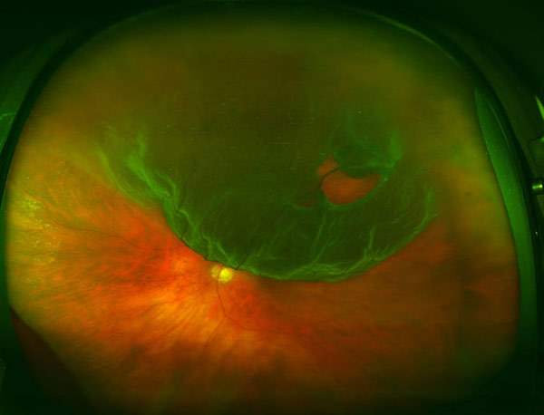
Retinal Detachment Caused by a Retinal Defect: Rhegmatogenous Retinal Detachment
Excessive pulling, especially if concentrated to a small area, can cause the retina to tear. Fluid from the vitreous cavity can come in through the hole and accumulate under the retina. This type of retinal detachment has an excessively complicated name, a rhegmatogenous retinal detachment. Retinal tears leading to detachment is the most common form of retinal detachment leading to eye surgery. The link between retinal tears and detachments didn’t happen that long ago in medical history. There was no way to look into the eye until about 150 years ago. It took many years to develop methods that allowed examination of the entire retina. Once that happened people realized the retina could detach, and they saw the retina had tears, but thought the tears occurred because the retina was detached, not the other way around.
Excessive Leakage under the Retina: Exudative Retinal Detachment
A complicated net of blood vessels exists under the retina in a structure called the choroid. The retina has the highest need for oxygen in the body per gram of tissue. If the retina had enough blood vessels to supply this requirement we would have a difficult time seeing because of all the blood vessels. Instead the eye has a nice design feature. A dense network of blood vessels delivers oxygen to the retina from below. This layer is called the choroid, and the choroid has the highest blood flow per gram of tissue in the body. Problems can arise if the vessels in the choroid become excessively leaky. If you have ever injured your elbow or knee you know the joint can become swollen. The swelling is from fluid leaking from blood vessels. Imagine the choroid – if that becomes inflamed there are many vessels that could leak. Fluid produced in the choroid will make that layer swollen, but the fluid oozes up under the retina and accumulates there. In exudative detachments the retina is floats off of the back of the eye.
Vitreous Retina Macula Consultants of New York
110 Lafayette St, Suite 502
New York, NY 10013
Tel.: (212) 234-3367
(212) 861-9797
Fax.: (212) 628-0698
Web Address https://www.vrmny.com/
Our location on the map: https://goo.gl/maps/QT1BMFr3djLXn6427
Nearby Locations:
Lower Manhattan | Little Italy | Chinatown | Civic Center | Tribeca | SoHo
10013 | 10012 | 10007 | 10002
Working Hours:
Monday-Friday: 8am – 5pm
Payment: cash, check, credit cards.
Our social links:
https://www.facebook.com/vrmny
https://twitter.com/vrmnyc
https://www.linkedin.com/company/vitreous-retina-macula-consultants-of-new-york
https://instagram.com/vrm.ny
https://www.youtube.com/channel/UCde-Yz2MUq8r6BmEAInNLrQ
https://www.yelp.com/biz/vitreous-retina-macula-consultants-of-new-york-new-york-4
https://vrmny.tumblr.com/
https://www.pinterest.com/vrmnyc
View other locations Vitreous Retina Macula Consultants of New York has been mentioned
http://www.yellowpages.com/info-571904193/v/
http://ablocal.com/us/new-york-ny/LX13924492-vitreous-retina-macula-consultants-of-new-york/
https://newyork.yalwa.com/ID_140802032/Vitreous-Retina-Macula-Consultants-of-New-York.html
https://www.2findlocal.com/b/14717613/vitreous-retina-macula-consultants-of-new-york-new-york-ny
https://www.americantowns.com/yp/vitreous-retina-macula-consultants-of-new-york-brooklyn-heights-ny-yx82579408.html